


Collaborating to Find a Cure for Blindness
For patients who find themselves slowly beginning to lose vision, today’s doctors have gene replacement therapy as a treatment option. However, there’s a small window of time when the therapy will work best, meaning if a patient isn’t seen soon enough, vision cannot be restored.
Such is the case with several diseases of the retina. But two researchers whose work has been funded by loyal donors hope to change that.
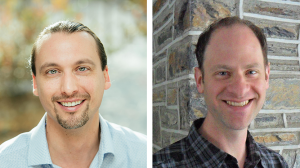
Greg Field, PhD, and Jeremy Kay, PhD, are each working on research related to the retina that could not only change the lives of people facing blinding disease but also potentially help people facing neural degenerative diseases such as Parkinson’s disease, Alzheimer’s disease, and ALS.
Up until now, Field and Kay have worked separately on projects related to the retina. By working together, they hope to advance the ability to treat several types of blinding diseases, such as retinitis pigmentosa, glaucoma, and macular degeneration. “This is so valuable because nearly all blinding diseases are diseases of the retina,” says Field, an assistant professor of neurobiology. “Studying and understanding degeneration of the retina is our best bet at curing blindness.
“The genetic basis of many kinds of retinal degeneration have a lot of similarity to the genetic basis of a variety of neurodegenerative diseases,” he adds.
Kay, assistant professor of neurobiology and ophthalmology, has been working to understand the development of the retina. He and his colleagues are experts in identifying the structure and the building blocks of the retina. Field’s lab is focused on understanding how the retina encodes everything that the eye sees and how it transmits that information to the brain.
“Those are two sides of the same coin,” Field says. “If the function (of the retina) changes, it’s hard to know what that means if you don’t know how the structure changes. Correspondingly, if you see structural changes, it’s not obvious what that means for the function. So, putting those two things together really gives us a complete picture of retinal disease progression.”
Kay’s lab previously made an unexpected finding about the genetic basis of retinal degenerative disease. They discovered that the degeneration originates in a previously unappreciated variant of one particular gene called CRB1. With current Holland-Trice funding, Kay plans to use a new method developed by his lab to look more closely at all of the known retinal disease genes with the hope of determining what about the genes leads to degeneration.
“With our new method we can interrogate that a little more closely and see if there might be some other hidden biology that might be important for treating the disease,” Kay says. “The method can be applied to any disease. The goal is to pilot it with retinal diseases, and once we’ve shown that it works there, we can then use it on neurodegenerative disease genes or intellectual disability disease genes.”
He hopes that the version of the gene that his lab discovered could be turned into gene replacement therapy with the help of Field and his lab.
“If we understand the genetic basis of the disease, we understand how it starts, and then maybe you can stop it by replacing the piece that’s broken,” Kay says. “We think we have a way of finding new pieces that are broken. But to use that appropriately, you need the information that Greg’s lab is generating.”
In degenerative diseases there are cells in the neural tissue that are sick and dying. This in turn causes healthy neurons to find other neurons to communicate with, and the brain and the retina start to rewire. However, it’s not clear to researchers whether this rewiring can be reversed simply by curing the sick cells—essentially replacing the missing or defective genes.
Using mice engineered with a genetic defect, Field and his colleagues have found that retinal degenerative disease can be cured; however, if the cure isn’t introduced early enough, the defective wiring retains its abnormal properties.
“If we intervene a little earlier, we see that the retinal cells that are rewired actually go back to their normal condition,” Field says. “They sort of un-rewire. What the Trice funding is allowing us to do is to figure out the genetic switches that are either allowing or not allowing rewiring to occur.”
He adds: “If we could figure out a secondary therapy, whether that’s a drug or a secondary gene therapy and stimulate these rewired circuits to go back to normal, that would be a huge advance.”
Field says they can do this in the retina first because more is known about how the retina works than any other part of the brain.
“The retina is the only part of the brain that doctors can directly look at without drilling a hole in your head or putting you in some kind of big magnet,” he says. “There’s a lot of hope that observing early changes in the retina can be useful for diagnostics of various kinds of neural degenerative diseases. This is a place to invest research dollars that not only pays dividends in helping people see, but I think it’s very likely to pay dividends in curing other brain diseases.”
“The retina is the only part of the brain that doctors can directly look at without drilling a hole in your head or putting you in some kind of big magnet,” he says. “There’s a lot of hope that observing early changes in the retina can be useful for diagnostics of various kinds of neural degenerative diseases. This is a place to invest research dollars that not only pays dividends in helping people see, but I think it’s very likely to pay dividends in curing other brain diseases.”
Greg Field, PhD
Both researchers are grateful for the funding they have received from philanthropy. Without it, they say it would have been difficult for their projects to move forward. The findings from Kay’s Holland-Trice Scholars funded work served as the key preliminary data for an NIH R01 application that was recently funded, yielding $750,000 in direct costs from the National Eye Institute.
Initial results from Field’s project have been accepted for a presentation at the annual meeting for the Association for Research in Vision and Ophthalmology. He also anticipates publishing findings in late 2020 or early 2021 and using the data to support an application to the National Eye Institute for additional external funding.
Greg Field, PhD
“The retina is the only part of the brain that doctors can directly look at without drilling a hole in your head or putting you in some kind of big magnet,” he says. “There’s a lot of hope that observing early changes in the retina can be useful for diagnostics of various kinds of neural degenerative diseases. This is a place to invest research dollars that not only pays dividends in helping people see, but I think it’s very likely to pay dividends in curing other brain diseases.”
Both researchers are grateful for the funding they have received from philanthropy. Without it, they say it would have been difficult for their projects to move forward. The findings from Kay’s Holland-Trice Scholars funded work served as the key preliminary data for an NIH R01 application that was recently funded, yielding $750,000 in direct costs from the National Eye Institute.
Initial results from Field’s project have been accepted for a presentation at the annual meeting for the Association for Research in Vision and Ophthalmology. He also anticipates publishing findings in late 2020 or early 2021 and using the data to support an application to the National Eye Institute for additional external funding.